Lymphatic tissue. Lymphoid tissue and its role in our resistance to disease
Introduction
Immunity (from the Latin immunitas - to free from something) is a physiological function that causes the body's immunity to foreign antigens. Human immunity makes him immune to many bacteria, viruses, fungi, worms, protozoa, and various animal poisons. In addition, the immune system protects the body from cancer cells.
The task of the immune system is to recognize and destroy all foreign structures. Upon contact with a foreign structure, cells of the immune system trigger an immune response, which leads to the removal of the foreign antigen from the body.
The function of immunity is ensured by the work of the body's immune system, which includes various types of organs and cells.
The anatomy of the immune system is extremely heterogeneous. In general, cells and humoral factors of the immune system are present in almost all organs and tissues of the body. The exception is some parts of the eyes, testicles in men, the thyroid gland, and the brain - these organs are protected from the immune system by a tissue barrier, which is necessary for their normal functioning.
In general, the functioning of the immune system is ensured by two types of factors: cellular and humoral (that is, liquid). Cells of the immune system (various types of leukocytes) circulate in the blood and pass into tissues, carrying out constant surveillance of the antigenic composition of tissues. In addition, a large number of different antibodies (humoral, fluid factors) circulate in the blood, which are also capable of recognizing and destroying foreign structures.
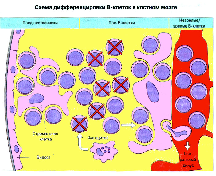
In the architecture of the immune system, we distinguish between central and peripheral structures. The central organs of the immune system are the bone marrow and the thymus (thymus gland).
The peripheral organs of the immune system are represented by the lymph nodes, spleen and lymphoid tissue (such tissue is located, for example, in the palatine tonsils, at the root of the tongue, on the posterior wall of the nasopharynx, in the intestines).
Since lymphoid tissue is located scattered throughout the body, in various structures of organs of all systems, generalizing knowledge about it is of interest, therefore the purpose of this abstract is to analyze and combine all the studied material about lymphoid tissue distributed throughout the body.
Lymphoid tissue of the mucous membranes, definition, functions, structure
Lymphoid tissue (anat. lympha, from lat. lympha pure water, moisture + Greek -eidзs similar) - a complex of lymphocytes and macrophages located in the cellular fibrous reticular stroma; constitutes the functioning parenchyma of central and peripheral lymphoid organs.
In addition to the mass of lymphoid tissue encapsulated in the spleen and lymph nodes, the body contains a significant amount of “free” lymphoid tissue not enclosed in a connective tissue capsule, which is localized in the walls of the gastrointestinal, respiratory and urogenital tracts and serves as protection against infection.
It is designated as lymphoid tissue associated with mucous membranes. In humans, these are the lingual, palatine and pharyngeal tonsils and Peyer's patches of the small intestine, the appendix.
The main effector mechanism of the immune response is the secretion and transport of secretory antibodies of the IgA class (sIgA) directly on the surface of its epithelium. It is not surprising that most of the lymphoid tissue is present in the mucous membranes and is especially abundant in the intestines, since it is mainly through the mucous membranes that antigens from the outside penetrate. For the same reason, IgA antibodies are present in the body in the greatest quantity relative to other antibody isotypes. Lymphoid tissue associated with mucous membranes, the protective effect of which is based on the production of IgA, is often abbreviated MALT (mucosal-associated lymphoid tissue). There is an assumption that mucous membrane-associated lymphoid tissue (MALT) forms a special secretory system in which cells that synthesize IgA and IgE circulate.
Once in the intestine, the antigen penetrates Peyer's patches through specialized epithelial cells and stimulates antigen-reactive lymphocytes. After activation, they pass with lymph through the mesenteric lymph nodes, enter the thoracic duct, then into the blood and into the lamina propria, where they turn into cells that produce IgA, and as a result of such widespread distribution, they protect a large area of the intestine by synthesizing protective antibodies. Similar cells are also concentrated in the lymphoid tissue of the lung and in other mucous membranes, apparently with the help of homing receptors similar to the MEL-14-positive receptors of the high endothelium of the lymph nodes. Thus, the migration of lymphocytes from lymphoid tissue to the blood and back is regulated by homing receptors located on the surface of high endothelial cells in postcapillary venules.
In almost all cases, lymphoid tissue is represented by two formations: lymph nodes and surrounding clusters of lymphocytes.
Lymph nodules (follicles) - zone of B lymphocytes
As in the follicles of the lymph nodes, the following areas can be distinguished in the lymph nodes (1) of the mucous membranes:
a) reactive center, including 3 zones:
Dark (where stimulated B cells - centroblasts - are in a state of mutagenesis),
Light basal (where the selection of centrocytes - products of mutagenesis) occurs,
Light apical (where cells intensively divide - B-immunoblasts - selected by the degree of affinity for the antigen);
b) as well as the surrounding crown (where differentiation of cells formed from B-immunoblasts, proplasmocytes and memory B-cells occurs).

Parafollicular accumulations of lymphoid tissue - T-zone
Parafollicular accumulations of lymphocytes (2) are an analogue of the paracortical zone of lymph nodes.
This means that these clusters are formed by T cells (representing the T zone), i.e. contain T-immunoblasts, memory T-cells and mature activated T-lymphocytes (killer T-cells, helper T-cells and possibly suppressor T-cells).
Non-lymphoid elements
Despite the similarity in the organization of lymphoid elements, there are differences from lymph nodes in relation to non-lymphoid components:
Firstly, the stroma is represented by reticular rather than loose connective tissue;
Secondly, the processing of antigens (by macrophages) and their presentation to lymphocytes (antigen-presenting cells) occurs mainly in the epithelium, which is associated with a different (than in the lymph nodes) localization of the cells involved in these processes.
Now let’s look separately at the various accumulations of lymphoid tissue.
One of the types of connective tissue in which the system of macrophages and lymphocytes is located is called lymphoid. It can be presented in the form of separate organs, or it can simply be a functioning part of the body. Lymphoid tissue is found in organs such as the bone marrow and spleen, lymph nodes and thymus. In them it is a functioning parenchyma.
In the mucous membrane of some organs there are also accumulations of lymphoid tissue - bronchi, urinary tract, kidneys, intestines and others.

Functions
In all protective reactions without exception, lymphoid tissue takes the main part. The lymphocytes, macrophages and blasts contained in it, plasma cells, mast cells and leukocytes protect the body from the invasion of foreign cells and remove damaged cells of the body itself. The lymph nodes and intestinal (lymphoid) tissue are responsible for the formation of cells of the immune system.
If a bacteria or virus enters through damaged skin, a defense reaction is activated in the lymph node closest to the site of penetration, lymphoid cells and macrophages are released, which move along with lymph and blood to the place where the “stranger” is found. In the event of a mass attack, when the forces of one lymph node cannot be dealt with, the entire immune system is activated.
Structure
Lymphoid tissue most often consists of free cells supported in a network of reticular fibers. The network can be denser in composition (forms a dense tissue) or loose (with spaces where free cells can move freely). The fibers themselves are formed from type III collagen.

Places of congestion
Large accumulations of lymphoid tissue are located in places where foreign organisms are most likely to enter. The tonsils, familiar to everyone, are the lymphoid tissue of the pharynx, located on the border with the oral cavity. They are pharyngeal, palatal, tubal and laryngeal. The totality of all tonsils and areas is the lymphoid tissue of the nasopharynx.
Its function is very important for our health, because it neutralizes germs entering through the mouth and nose. And together with organs containing lymphoid tissue, it ensures the formation of the required number of lymphocytes for the whole organism.
Among other things, the lymphoid tissue in the throat interacts with the endocrine glands (adrenal glands, thyroid gland, thymus, pancreas), forming a close connection “pituitary gland - adrenal cortex - lymphatic tissue” until the child reaches puberty.
What is hypertrophy
A child from three to ten years old may develop hypertrophy of the lymphoid tissue of the tonsils, but its functioning is not impaired. Only with the beginning of puberty does the hypertrophied tissue begin to decrease.
It is not known exactly what this process is associated with, but the presumed causes are inflammation of the pharynx or infection, various endocrine disorders. Hypertrophy can lead to frequent inflammation or pathological changes in the ears, nose, and larynx.

If nasal breathing is impaired, ventilation of the lungs is weakened. Later, this leads to a change in the composition of the blood - hemoglobin and the number of red blood cells decrease, and leukocytes increase in number. Then the functions of the gastrointestinal tract, thyroid gland, and adrenal glands begin to be disrupted. Violation of all processes leads to delays in the growth and sexual development of the child.
What is hyperplasia
The term "hyperplasia" comes to us from the Greek language and means over-education. At its core, this is a pathology in which cells begin to multiply intensively, increasing the volume of tissue.
- Infectious. The immune response to any infection leads to the rapid production of lymphocytes and macrophages, which causes the proliferation of lymphoid tissue.
- Reactive. Bacteria and microbes enter the lymph node, where their waste products and toxins they secrete accumulate, causing, in turn, the active release of macrophage cells.
- Malignant. Any cells of the lymph node can be involved in this pathological process, which leads to a change in its size, shape and structure.
Lymphoid tissue is one of the most important components of our body’s immune system. It helps prevent many diseases even before infection gets inside along with food and air. It also performs other functions, the mechanism of which has not been fully studied.

Sometimes the lymphoid tissue becomes inflamed, and diseases such as appendicitis, tonsillitis and many others appear (depending on the location of the lymphoid tissue). Very often in such cases, doctors resort to surgical treatment methods, in other words, they remove the affected area or organ. Since all the functions of lymphoid formations have not been fully studied, it cannot be said 100% that such removal does not harm the human body.
Moisture + Greek -eidēs similar) a complex of lymphocytes and macrophages located in the cellular fibrous reticular stroma; constitutes the functioning parenchyma of lymphoid organs. Lymphoid organs, which are organs of immunogenesis, include the thymus gland (thymus gland) ,
The lymph nodes ,
spleen (Spleen) ,
lymphoid elements of the bone marrow and accumulations of L.t. in the walls of the gastrointestinal tract, respiratory and urinary tract. The basis of L.t. consist of reticular fibers and reticular cells, forming a network with cells of various sizes. In the loops of this network there are cells of the lymphoid series (small, medium and large lymphocytes, plasma cells, young cells - blasts), macrophages, as well as a small number of leukocytes, mast cells. Reticular cells are formed from mesenchyme, and lymphoid cells are formed from bone marrow stem cells. Cells of the lymphoid series, among which two populations are distinguished - T- and B-lymphocytes, move with blood and lymph. Together with macrophages, they participate in immune responses against genetically foreign substances (see Immunity) .
The structure of L.t., the topography of its structural elements in various organs of the immune system has its own characteristics. In the central organs of immunogenesis L.t. is in functional unity with other tissues, for example in the bone marrow - with myeloid tissue, in the thymus gland - with epithelial tissue. In the peripheral organs of the immune system, for example in the walls of the gastrointestinal tract, respiratory and urinary tract, depending on the degree of maturity and functional state of L.t. is in various qualitative states - from single lymphocytes and diffusely located lymphoid tissue to lymphoid nodules with reproduction centers, the presence of which indicates the high immune activity of the body. The largest number of lymphoid nodules, including those with reproductive centers, are found in the tonsils, lymphoid plaques, spleen, walls of the appendix, stomach, small and large intestines, and lymph nodes in children and adolescents. In addition to accumulations, L. t. in the form of a rare, thin, seemingly protective layer of cells of the lymphoid series is located under the epithelial cover of the respiratory and urinary tracts and the gastrointestinal tract. In the spleen it forms lymphoid muffs around arterial vessels. As the body ages, the amount of Lt decreases. and lymphoid nodules in the organs of the immune system. In inflammatory processes and activation of immune reactions, both primary and secondary (see Immunopathology) ,
Reactive lymph nodes are observed. L. t. is affected by hemoblastoses (Hemoblastosis) ,
histiocytosis (Histiocytosis X) X, Lymphogranulomatosis ,
malignant lymphomas, paraproteinemic hemoblastoses (Paraproteinemic hemoblastoses) .
Bibliography: Sapin M.R. Immune structures of the digestive system, p. 123, M., 1987; aka, Principles of organization and structural patterns of organs of the human immune system, . anat., histol. and embryol., t. 92, no. 2, p. 5, bibliogr.
1. Small medical encyclopedia. - M.: Medical encyclopedia. 1991-96 2. First aid. - M.: Great Russian Encyclopedia. 1994 3. Encyclopedic Dictionary of Medical Terms. - M.: Soviet Encyclopedia. - 1982-1984.
See what “Lymphoid tissue” is in other dictionaries:
Lymphoid tissue of the walls of the respiratory and digestive systems- An accumulation of lymphoid tissue containing, against a background of diffusely located cellular elements, follicles, which are a denser (nodular) accumulation of cells, called tonsils (tonsillae). Tonsils, located in the initial... ... Atlas of Human Anatomy
- (textus, LNH) a system of cells and non-cellular structures united by a common function, structure and (or) origin. Granulation tissue (granulationes; synonym: granulation, T. granular) connective T., formed during the healing of tissue defects ... Medical encyclopedia
- (lat. cambium exchange, change) general name for T., in which intensive cell division occurs (for example, lymphoid tissue, intestinal epithelium) ... Large medical dictionary
Tissue that produces cells that carry out the body's defense reactions - lymphocytes and plasma cells. It is present in the body in the form of many discrete formations (for example, lymph nodes, tonsils, thymus gland and ... ... Medical terms
- (t. lymphadenoideus) see Lymphoid tissue... Large medical dictionary
- (t. lymphoreticularis) see Lymphoid tissue... Large medical dictionary
LYMPHOID TISSUE- (lymphoid tissue) tissue that produces cells that carry out the body’s protective reactions - lymphocytes and plasma cells. It is present in the body in the form of many discrete formations (for example, lymph nodes, tonsils, thymus... Explanatory dictionary of medicine
- (t. lymphoideus; lymph + Greek. eides similar; synonym: T. lymphadenoid, T. lymphoreticular) reticular T. with a large number of lymphocytes; forms the parenchyma of the lymph nodes, spleen, tonsils, thymus, lamina propria... ... Large medical dictionary
Lymphatic system- is part of the cardiovascular system and complements the venous system, takes part in metabolism, cleanses cells and tissues. It consists of lymphatic pathways that perform transport functions, and organs of the immune system that perform the functions... ... Atlas of Human Anatomy
The lymph nodes- (nodi lymphatici) the most numerous organs of the immune system. In the human body, their number reaches 500. All of them are located in the path of the lymph flow and, by contracting, contribute to its further advancement. Their main function is... ... Atlas of Human Anatomy
Immune system organs- The immune system provides immune protection to the body through the cellular elements of the immune system, which are lymphocytes and plasma cells. The immune system is made up of the lymph nodes, spleen, bone marrow, thymus gland, or thymus... Atlas of Human Anatomy
Lymphoid tissue is very sensitive to external and internal influences. As the body ages, the amount of Lt decreases. and lymphoid nodules in the organs of the immune system.
Lymphoid tissue (synonym lymphatic tissue) is a collective term for the structures in which lymphocytes are formed. Human lymphoid tissue makes up about 1% of body weight and is one of the most important components of lymphoid organs.
What is Hypertrophy of pharyngeal lymphoid tissue -
One of the main functions of lymphoid organs is their participation in the processes of hematopoiesis (lymphopoiesis). This ability of lymphocytes is associated with an important function of lymphoid tissue - its participation in the body’s defense reactions. Hormones of the adrenal cortex have a great influence on the degree of development of lymphoid tissue. Insufficient function of the adrenal cortex causes the proliferation of lymphoid tissue. The administration of adrenal hormones leads to degeneration of lymphoid tissue and death of lymphocytes.

The structure and role of lymphoid tissue in the activity of the immune system
The structure of L.t., the topography of its structural elements in various organs of the immune system has its own characteristics. In the central organs of immunogenesis L.t. is in functional unity with other tissues, for example in the bone marrow - with myeloid tissue, in the thymus gland - with epithelial tissue. In addition to accumulations, L. t. in the form of a rare, thin, seemingly protective layer of cells of the lymphoid series is located under the epithelial cover of the respiratory and urinary tracts and the gastrointestinal tract.
Lymphoid tissue of the mucous membranes: introduction
Lymphoid organs are classified as either primary (central) or secondary organs. Thus, lymphocytes belong to the category of cells that are widely distributed in the body. Lymphoid tissue is a type of connective tissue that is characterized by a high content of lymphocytes.

In most lymphoid organs, fibroblast-like reticular cells form these fibers, on which their numerous processes are located. Nodular lymphoid tissue is formed by spherical clusters of lymphocytes; these are the so-called lymphoid nodules, or lymphoid follicles, containing predominantly B-lymphocytes. Lymphoid tissue associated with mucous membranes, the protective effect of which is based on the production of IgA, is often abbreviated MALT (mucosal-associated lymphoid tissue).
The lingual tonsil consists of accumulations of lymphoid tissue - lymphoid nodules, the number of which (80-90) is greatest in childhood, adolescence and young adulthood. By the time of birth, the number of lymphoid nodules in the developing tonsil increases markedly. Reproduction centers in lymphoid nodules appear soon after birth (in the 1st month of life). Subsequently, their number increases until adolescence.
The structure of lymphoid tissue. Histology, functions
The branches of the right and left lingual arteries, as well as, in rare cases, branches of the facial artery approach the lingual tonsil. Trabeculae (septa) extend from this plate in the medial direction into the lymphoid tissue of the organ, which, if well expressed, divide the tonsil into lobules.
In a 5-month-old fetus, the tonsil is represented by an accumulation of lymphoid tissue up to 2-3 mm in size. During this period, epithelial strands begin to grow into the developing tonsil - future crypts are formed. On the surface of the folds in children, numerous small tubercles are visible, in the depths of which there are accumulations of lymphoid tissue - lymphoid nodules.
Under the epithelial cover in the diffuse lymphoid tissue there are lymphoid nodules of the pharyngeal tonsil with a diameter of up to 0.8 mm, most of which have reproduction centers. The pharyngeal tonsil is formed in the 3-4th month of intrauterine life in the thickness of the developing mucous membrane of the nasal part of the pharynx.
By the end of the year, its length reaches 12 mm and width - 6-10 mm. lymphoid nodules in the tonsil appear in the 1st year of life. After 30 years, the size of the pharyngeal tonsil gradually decreases. Age-related involution of the tubal tonsil begins in adolescence and young adulthood. It is usually observed in children aged 3-10 years. Hypertrophied lymphoid tissue undergoes physiological involution and decreases during puberty.
While maintaining its function, hypertrophied lymphoid tissue can, however, cause pathological changes in the nose, ears and larynx. Hypertrophy of the palatine tonsils is often combined with hypertrophy of the entire pharyngeal lymphoid ring, especially with hypertrophy of the pharyngeal tonsil. During puberty, the adenoids undergo reverse development, but the resulting complications remain and often lead to disability. Indirect signs of adenoids are also hypertrophy of the palatine tonsils and lymphoid elements on the posterior wall of the pharynx.
Hypertrophy of lymphoid tissue in response to an infectious disease leads to an increase in inflammatory processes in the pharynx. In the thickness of the tonsil there are rounded dense accumulations of lymphoid tissue - lymphoid nodules of the tonsil. Areas of lymphoid tissue are found in the mucous membrane of some organs (bronchi, urinary tract, kidneys).