What can replace a colonoscopy of the intestine. How to examine the intestines without colonoscopy - possible diagnostic methods
The number of cancer patients is increasing every year. Colon cancer ranks third in the number of deaths. Oncology affects people from 45 years old, the younger generation often gets sick. Persons with oncological heredity should carry out prophylaxis every six months in a medical institution. It is necessary to check the intestines if there is a genetic predisposition to neoplasms.
Patients are afraid of this research method (colonoscopy) and try to find alternative methods. The presented methods are informative and will help to examine the organ. Colonoscopy research method is unpleasant, requires long and special preparation. Use other methods of diagnosing bowel diseases that come to the aid of the patient in addition to colonoscopy. The peculiarity of these methods is that when a pathology is detected in the intestine, it is impossible to take a piece of material for analysis. No analogue can replace a full-fledged study.
Before any diagnostic method, you should not eat food and drink plenty of fluids. The results will be more reliable. Patients who have had a colonoscopy are looking for other alternative methods for diagnosing bowel disease. Any alternative method can exist separately. But in severe cases, colonoscopy is indispensable.
The procedure does not reveal all neoplasms. Colonography does not damage the mucosal wall. It is possible to study and consider the contours of the foci and the state of nearby organs. The procedure for a CT scan is similar to an x-ray. The apparatus takes several shots, performing a systematic storage in large numbers. Tomography without colonoscopy is not able to detect cancer in the first stage. For this method, you need to drink a solution or inject the composition. The technique is carried out longer than an x-ray, a person should be quietly in a lying position for a while, without moving.
Virtual tomography operates using a special computer program that analyzes the results of CT and is able to detect polyps, growths of more than 1 cm. This method is not used in any center, early detection of diseases with its use is excluded.
Ultrasound examination (ultrasound)
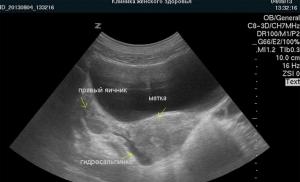
This diagnostic method is used instead of colonoscopy in some situations. There is a registration of sound waves reflected from the boundaries of tissues. The study will allow you to explore the lesion of the neoplasm. The device sees nodes ranging in size from 0.5-2 cm.
Two methods are used to examine the body. Ultrasound of the abdominal cavity, but in 20% of cases it is difficult to analyze the rectum due to the low filling of the bladder. An alternative ultrasound method is to examine the colon using a transducer that is inserted through the colon.
The indications are:
- constant stool retention;
- lack of stool management;
- the presence of blood in the feces;
- on palpation, formations in the rectum are felt;
- during the x-ray, a deviation of the organ was detected, with sigmoidoscopy, a change in the shape of the intestine was detected;
- colonoscopy showed cancer;
- for the diagnosis of pathology of the large intestine;
- the patient is at risk for oncology;
- a person is treated with clinical signs of a lesion of the digestive system.
Indications for the method: growth of formations in the intestine continues, an increase in the number of malignant formations, elimination of invasion of prostate cells into the intestinal region, examination of complications after tumor removal.
Irrigoscopy
The technique is able to study the intestine without the use of colonoscopy - to assess the location of neoplasms, tumors, their dimensions, shape and maneuverability. The method is carried out by introducing an enema of a barium solution with a bright substance. Then the doctor takes an x-ray. When barium sulfate is removed, air is introduced. Thanks to this, it is possible to examine the outlines of organs, detect fistulas, ulcers. It turns out to evaluate the structural features and functionality of the large intestine. The procedure is safe and painless.
It is carried out by doctors using conventional x-rays. You will first need to carry out preparatory manipulations:
- Perform bowel cleansing with an enema and a special drug.
- Do not shower before the procedure.
The examination is characterized by signs: discomfort and pain in the anus, a bloody mass is released from the anus during or after stool. The indication for the procedure will be long-term diarrhea, chronic stool retention, discharge from the anus of various etiologies, acute pain in the abdomen, flatulence. It is possible to detect an outward formation, but it is impossible to examine the structure and take a biopsy.
Capsule examination
This is an innovative diagnostic method. If the patient has individual characteristics of the intestine, and it is impossible to examine the diseased organ using standard methods, then this method of examination is used. A capsule 10 mm in size, 30 mm long, which is equipped with cameras, is swallowed by the patient. The device moves through the intestines, takes a picture and is brought out. Snapshots occur frequently - from 4-40 shots per second. It depends on the speed of movement. With the help of waves, information is transmitted to special specialized equipment.
The procedure takes 5-8 hours, painless. It is impossible to get infected with diseases, the capsule is sterile and disposable. It is prescribed for hidden bleeding, neoplasms and other pathologies. The procedure finds the cause of the disease in the intestines and gastrointestinal tract. Pretty comfortable for the patient. For example, it is possible to read a book, walk and watch TV.
Anoscopy
Using the method, up to 10 cm of the lower segment of the rectum is examined. A medical optical device with illumination is inserted into the intestine - an anoscope. Neoplasms, nodes, inflammations, polyps are determined. You can take a biopsy.
Sigmoidoscopy
Endoscopic diagnostic method. The procedure is carried out every five years. Examine only 30 cm of the intestine. You can take neoplasm tissue. Inaccurate diagnosis of diseases. After the procedure, a number of other methods are prescribed, many of which are more effective.
Hydrogen test
The procedure takes place within three hours. Every 30 minutes, the patient exhales into a special tube. They study a significant number of bacteria that enter the small intestine. The principle of operation of the method: bacteria do not allow liquid to penetrate into the intestinal mucosa in a normal amount, therefore defecation is disturbed. When there are common symptoms, the indicated method is needed:
- irritable bowel syndrome;
- sugar intolerance;
- the intestines do not absorb products (cow's milk, some fruits, honey);
- increased accumulation of bacterial flora;
- small production of juice for the function of digesting food;
- symptoms of altered and disturbed microflora (flatulence, diarrhea, constipation);
- evaluation of the effectiveness of the treatment of intestinal diseases that are associated with atrophy of the intestinal villi lining the walls.
Magnetic resonance imaging and colonography
MRI is an alternative to colonoscopy, but more expensive. This procedure is prescribed in addition to the examination. MR colonography is a procedure for examining the intestines for diseases. Two liters of liquid with a bright color are injected into the rectum, with the help of the device they look at the state of the organ in a three-dimensional image. The procedure takes an hour. Contraindication to the procedure of colonography - persons with kidney disease.
PET - positron emission tomography
The procedure lasts 1.5 hours. One hour the patient waits for the results. Radioactive sugar is used in the examination method. Enter intravenously. With its help, it is possible to diagnose oncological diseases, tumors. Pathological cells absorb the substance - it is easy to see their location.
This is from the field of nuclear medicine, created by using a special type of scanner and atoms to establish an assessment of the functioning of organs. The effectiveness of the method depends on the drug used.
The method is prescribed together with CT. The combination of PET results with CT images allows you to acquire detailed information about the location of radioactive elements. Determine the stages of oncology, check the function of blood flow or the functioning of organs. in the survey. They are needed to detect the initial stages; for an accurate diagnosis, one cannot do without the PET technique. If the disease is mild, the examination takes place with the help of palpation, tapping, examination and listening. Often diseases are determined by laboratory tests of urine, feces, blood. Replacing colonoscopy in some cases will be considered an underexamination.
Today, a number of alternative diagnostics have been developed that complement colonoscopy. It is impossible to completely replace the method, alternative methods are not so accurate. Some are used only in a narrow specialty, others are not allowed and contraindicated due to staining substances, but the patient needs to go through a colonoscope. This device is the only way to diagnose diseases, take samples for analysis and prescribe the right treatment.
At the diagnostic stage with the method of colonoscopy, the possibility of ridding the body of feces, growths and other benign polyps is considered. Helps to cleanse the intestinal spaces, the function of which is complicated by accumulated toxins. Research is also significant in the field of premature testing of oncological diseases, which gives the possibility of healing at an early stage, further complete cure of the disease.
Alternative methods - methods of the preparatory stage before colonoscopy, can help and detect diseases, but will not replace colonoscopy.
Those who have already experienced this unpleasant procedure are interested in how to check the intestines without a colonoscopy? After all, this diagnostic method not only gives the patient a lot of discomfort, but also requires long and complex preparation. The effectiveness of the procedure is invaluable, but still patients prefer to look for an alternative. Modern medicine offers them other methods of diagnosis, which in some cases can really be replaced by colonoscopy.
A special flexible tube with a camera located at its end is inserted into the examination process through the rectum. Thus, it is possible not only to examine the large intestine, but also to remove fecal stones or existing polyps from its walls. Colonoscopy is not used to examine patients with heart, lung or kidney failure, with exacerbation of intestinal infection, bleeding disorders, colitis, and peritonitis.
In addition to the fact that the procedure itself gives the patient a lot of discomfort and discomfort, you still need to carefully prepare for it.
During the day before the colonoscopy, the patient needs to follow a liquid diet, take laxatives and do enemas. All this is necessary to completely cleanse the intestines.
How you can check the intestines and how to replace this or that procedure, the doctor must decide. Making a choice in favor of colonoscopy, the doctor will tell the patient about the advantages and disadvantages of such a diagnosis.
The positives include:- Today, this method is considered the most effective and reliable.
- Due to the wide visualization, it is possible to fully assess the condition of the organ.
- During the procedure, it is possible, without resorting to surgery, to remove polyps and stop bleeding.
- Diagnostics takes no more than 30 minutes.
- The doctor may take tissue samples for a biopsy.
- Difficult and lengthy preparation for the examination. It is equally important to prepare mentally.
- The patient experiences discomfort during the procedure. Some people need to use sedatives or even general anesthesia.
Only a doctor can decide whether it is possible to check the intestines, except for colonoscopy, by other methods, after evaluating the general condition of the patient, a preliminary diagnosis and possible complications. Many are interested in what can replace a colonoscopy? Consider the most common methods for examining the intestine.
If during the examination the doctor suspects that the patient has any pathology of the intestine, he will prescribe a detailed examination. Colonoscopy has always been considered the gold standard.

But there are moments that do not allow its use (not all medical institutions have the necessary equipment and there are a number of contraindications to its implementation, including pregnancy, ulcerative colitis, some features of the patient's body, Crohn's disease or the presence of a disease such as diverticulitis in remission) .
In such cases, other methods of examination of the intestine may be used instead of colonoscopy.
Hydrogen test
The procedure consists in the fact that the patient needs to sit in one position for several hours. Every half an hour, he must breathe into a device that calculates how much hydrogen is released within the small intestine by bacteria.
The fact is that microorganisms disrupt the absorption of fluid by the intestinal mucosa, as a result of which the patient suffers from bloating and diarrhea. At the same time, carbohydrates are quickly decomposed into their constituent parts, and hydrogen is brought out during respiration.
Sigmoidoscopy and anoscopy
Sigmoidoscopy.
Examination of the intestine without colonoscopy by this method requires the use of a special apparatus of the rectoscope (a plastic device that has a depth scale and illumination). The procedure, as an analogue of colonoscopy, is prescribed in case of bleeding or pain in the sphincter.
A special tube is inserted into the rectum to a depth of no more than 35 cm and makes it possible to examine the sigmoid colon. During the diagnosis, the doctor can comprehensively assess the condition of the mucous membranes, blood vessels, measure the diameter of the lumen, detect scars, cracks and polyps.
For a better view, air pumping is used. Since sigmoidoscopy causes significant discomfort to a person, it is often performed under general anesthesia.
Anoscopy.
The procedure is very similar to sigmoidoscopy, but in this case, the tube is inserted no more than 12 cm. An anoscope can take tissue for analysis.
Irrigoscopy
This method allows you to replace. During the diagnosis, the doctor will conduct a visual examination of the intestinal walls, assess their stretching. Within 3 days before the procedure, the patient must follow a special diet. Additionally, cleansing with enemas is carried out.
The intestinal cavity is filled with a barium mixture. Thanks to this solution, the folds are straightened, and part of the organ is stained with a contrast agent to make the pictures better. This method of examination will be of interest to those who are interested in how to check the intestines for oncology without colonoscopy?
 This technique is an excellent alternative to colonoscopy of the intestine and is recommended for everyone who, for one reason or another, is unable to undergo it. For this, a special mini camera equipped with a light source is used. From above it is covered with a shell.
The patient needs to swallow this capsule and put on a cuff in which a recording device is placed that records the necessary indicators obtained directly from the capsule. The convenience of this diagnostic method lies in the fact that the patient does not need to be distracted from his daily activities. Passing through the entire digestive tract, the tablet will take photographs and various measurements to identify possible diseases of the digestive tract. Many consider it a worthy alternative to colonoscopy.
After 6-8 hours, the tablet with the camera will naturally leave the body, and the doctor will have all the information he needs. The only disadvantage of such a procedure is the inability to take a tissue sample for analysis.
Ultrasound, MRI and CT
These types of diagnostics can also be used as an alternative method of colonoscopy:
- ultrasound. Unfortunately, this method is not possible to qualitatively examine the patient. Ultrasound does not make it possible to determine oncological ailments at the initial stage of their development. With the help of ultrasound, the presence of metastases in the case of bowel cancer is checked.
- MRI. With the help of magnetic resonance imaging, the intestines can be examined for the presence of large neoplasms and foreign objects. If you first enter the substance gadolinium, you can make a diagnosis of polyps.
- CT. When choosing a way to examine the intestines without colonoscopy, sometimes they stop at computed tomography. However, it cannot give a complete picture in the case of cancerous tumors, since it is quite difficult to assess the state of small neoplasms. At the same time, CT provides a clear image of the organ and does not damage the mucous membranes and skin.
Non-instrumental diagnostic methods
Gastroenterologists believe that if diseases are caused by malnutrition and do not have a serious etiology, then non-instrumental research options are allowed.
In such cases, palpation of the peritoneum, tapping, listening and visual inspection of the abdomen are performed. Some diseases can be identified by hollowness or, conversely, swelling, the place of localization of unpleasant sensations and their nature (dull, acute, etc.).
In such cases, a history taking, urine and blood tests, liver tests, and a pancreatic examination may be sufficient to make a diagnosis. Proctologists, in turn, check the condition of the intestine using the anal-finger method, when the elasticity of its walls and mucous membranes is examined.
Invasive and minimally invasive diagnostics is often hampered by multiple contraindications, as well as the complexity of the manipulation: age-related features, the need for anesthesia, side effects during the administration of anesthesia. Classical colonoscopy is an adequate and only method for assessing the condition of the intestine, however, if it is impossible to perform, alternative methods can be used.
Alternative to colonoscopy
There are two types of palpation method:
- Surface. Atypical protrusions, locations of the painful focus are obvious to the doctor.
- Deep. Increased pressure and negative reaction of the patient to the examination of the abdominal space (normally, patients do not react to palpation in this area).
In addition to palpation, the doctor may prescribe other studies that may indirectly indicate the development of pathological changes in the intestinal mucosa:
- blood, urine, mucous component tests;
- analysis of feces for dysbacteriosis, eggworm, occult blood;
- general and detailed blood test.
In addition to colonoscopy, the most effective research method is capsule diagnostics. Compared to colonoscopy, the method is painless and not associated with complications. Patients only need to swallow a special capsule, inside of which a microscopic camera is implanted. Visualization occurs from the moment of ingestion to complete elimination from the body in a natural way. Capsule diagnostics requires special preparation and compliance with the doctor's recommendations.
On a note! Additionally, doctors resort to consultations of specialized specialists, carry out:
- ultrasound examination,
- bowel X-ray,
- computed tomography or MRI diagnostics.
The main analogues are research methods without colonoscopy for oncology and other diseases
In addition to colonoscopy, there are several effective methods for examining the intestinal cavities to exclude or differentiate one disease from another with a similar symptomatic course.
Alternative methods of examination are as follows:

It is difficult to unequivocally answer the question of the effectiveness of any alternative methods of colonoscopy. So, if a biopsy is necessary for suspected cancer or against the background of severe bleeding, including coagulation and the need to remove polyps, doctors again resort to traditional methods - endoscopic colonoscopy.
Modern methods of examination of the colon
 Colon examination can be performed by alternative methods, subject to the indications of the attending physician.
Given the anatomical proximity to the rectal lumen, the study is carried out in the following ways:
- Palpation of the rectal lumen. The study evaluates the condition of the mucous membranes of the anal sphincters, hemorrhoids, in some cases it allows to identify. Before the study, a cleansing enema is required. In case of insufficient information, other research methods are assigned.
- MRI diagnostics. An informative and high-precision method for studying soft tissues. The tomograph assesses the condition of the mucous membranes by layers, recognizes tumors less than 0.5 mm. The disadvantage of the method is the lack of guarantee of results in the study of the internal structures of the organ.
- CT scan. The procedure involves the study of the intestines by X-rays using a CT scanner. The effectiveness of the method is due to X-ray visualization of the smallest structures of the mucous epithelium, all fragments of the intestine with a pathological change.
- Sigmoidoscopy. A reliable method for studying any pathologies of the intestines, removed up to 30 cm from the anus. The main indications for carrying out are bloody discharge, pain during defecation. Sigmoidoscopy allows not only to assess the nature of the mucous structures, but also to collect histological material for further research.
- Anoscopy. Instrumental research method combined with palpation. With the help of anoscopy, a biopsy is available for further histology. As a preparation, a simple cleansing enema is suitable.
- Irrigoscopic examination. It involves taking x-rays in several projections with the obligatory introduction of a contrast agent.
- Sonography. The method is used for complaints of regular constipation and pathology of any origin. Echography is carried out in stages in which conditions are artificially created to improve the assessment of the state of the intestinal cavities. After emptying the intestines, the entire organ takes on its former forms.
Attention! Checking by alternative methods of examination of the colon is carried out as a differential diagnosis with chronic hemorrhoids, latent tumors near the rectal sphincter. With a doubtful diagnosis, traditional colonoscopy is usually resorted to.
Alternative to sigmoid colonoscopy
The sigmoid colon is the most important segment of the rectum, where absorption and distribution of nutrients throughout the body, the formation of feces takes place.
To determine the pathologies of the sigmoid colon, the following methods are used:
- Sigmoidoscopy. The accessibility of the method is determined by the distance of the possible study. With the help of sigmoidoscopy, the intestine is examined with a length of 25 cm.
- Irrigography. The study of the intestine with contrast is similar to other parts of the digestive tract.
If the diagnosis is unclear, CT diagnostics, MRI examinations are mandatory. In the absence of contraindications, they return to the "gold standards" of instrumental diagnostics - colonoscopy. For pain relief is now widely used.
The main differences between rectoscopy, anoscopy and colonoscopy in this video:
Latest Methods
Modern proctology offers patients various methods for examining various sections of the intestine in accordance with complaints. The choice of the method is due mainly to the indications for the study and the patient's complaints.
Differences between colonoscopy and videocolonoscopy
Video colonoscopy is a type of endoscopic colonoscopy in which a video of what is happening is made during the manipulation. During the manipulation, doctors have the opportunity to record the entire process, and then study fragments of particular interest.
With traditional colonoscopy, there is only an optical increase in the studied space with the possibility of photofixation.
Video colonoscopy is carried out similarly to traditional colonoscopy, only during operation a more modern colonoscope is used, equipped with the necessary equipment for video filming. The method is perceived by many clinicians as the "gold standard" in endoscopy.
How is it different from endoscopy?
The technical similarity of both procedures also determines their difference, which lies in the examined organ. Basically, a traditional colonoscopy is a specialized type of endoscopic examination of the bowel. With both procedures, it is possible to perform a biopsy, assess the condition of the mucosa, remove polyps and other non-oncogenic neoplasms, and also coagulate bleeding vessels.
Thus, there are no particular differences in procedures, except for the difference in the types of endoscopic equipment used. So, if it is necessary to carry out surgical manipulation, surgeons need an endoscope with two instrumental passages.
Replacement feasibility
Alternative diagnostic methods to colonoscopy are used under the condition of contraindications to the traditional examination. Given the need for anesthesia and the complexity of manipulation for some categories of patients, doctors resort to more gentle or completely non-invasive methods of studying the intestinal space.
The alternative to a full-fledged endoscopic examination is largely ineffective, which negatively affects the primary diagnosis of the patient. So, it is impossible for a doctor to conduct a full study of the clinical situation and assess the condition of the cavities of the intestinal segments, especially if surgical manipulations are necessary.
You can make an appointment with a doctor directly on our resource.
Be healthy and happy!
Those who have already experienced this procedure are looking for a way to check the intestines without a colonoscopy, since not only is the procedure itself quite unpleasant, but the preparatory stage before it takes a lot of time and effort. No one denies its effectiveness and efficiency, indispensability in terms of obtaining information, but a person tends to want to do without unpleasant sensations, especially if he knows about the existence of alternative methods. Modern research methods do offer other options for obtaining the necessary information, which in some cases allows them to replace colonoscopy.
On the procedure and explainability of the desire to replace it
Intestinal colonoscopy is performed by inserting a flexible tube with instruments and a camera at the end into the large intestine. When viewed from the walls of the intestine, the noticed polyps and fecal stones can be removed along the way. Warning that the procedure as a whole is quite tolerable, the proctologist does not tell the whole truth, but in some cases prescribes sedatives. This method is not applicable in case of hepatic, pulmonary, heart failure, peritonitis and colitis, bleeding disorders and acute intestinal infections.
In addition to the aesthetic ugliness of the procedure, there is also a preparatory period in which the patient spends 24 hours before the examination in the toilet or near it. This is due both to the liquid diet prescribed before the study, and to the laxatives and enemas prescribed to cleanse the bowel. If alternative methods can be dispensed with, patients prefer them. Colonoscopy is performed only in cases where the doctor needs complete and objective information.
Alternative Research Methods
In addition to colonoscopy, there are 7 instrumental methods for diagnosing the condition of the intestine. The only thing in which they are inferior to a colonoscopic study is that if negative phenomena are detected in the intestine, it is noted that it is impossible to take tissue from a problematic formation for analysis. Other methods of examining the intestine do not allow this, and if this kind of pathology is found, you will have to return to the intestine with special devices at the end. The examination of the proctologist is carried out by the following methods:
- CT scan;
- magnetic resonance imaging (MRI);
- irrigoscopy with barium;
- positron emission tomography (PET);
- capsule endoscopy.

Computed tomography is similar to an x-ray, but instead of one image, the tomograph takes them in layers, gradually producing images in large numbers. Computed tomography examination of the intestine without colonoscopy can not always detect cancer in the initial stage, which is always within the power of a proven method. For such a study, a contrast solution is drunk or an injection of the same substance is made. The procedure lasts much longer than the X-ray examination, and during this time the patient must lie motionless on the table.
Virtual tomography works using a program that processes CT results and can detect polyps larger than 1 cm, but this research method is not available in every medical center, and early diagnosis with its use is excluded. And if polyps are found, they will still have to be removed.

MRI is based on the use of magnets and radio waves, the energy of which is directed at the body and then returned in the form of reflected pulses. This method is based on the introduction of a drug with gadolinium, which behaves differently in diseased and healthy tissues, allowing the identification of polyps based on the decoding of the template into a detailed image using a computer program. This bowel examination is contraindicated in people with kidney disease.
PET uses a radioactive sugar, fluorodeoxyglucose, for research. The test allows you to explore the area around the anomaly, the condition of the lymph nodes and surrounding organs in the event that cancer has already been diagnosed, but does not provide tangible indications for direct diagnosis. To obtain complete information, the doctor needs to see a preliminary CT scan.

Ultrasound is rarely used, since it can only determine the stage of cancer germination or a fairly large tumor. It is most often used as an endorectal ultrasound for examining the rectum, using a special probe inserted into the immediate area of the examination.
Capsule endoscopy is applicable to the study of veins, muscular membranes and intestinal mucosa and is performed by swallowing a special capsule that takes pictures and transfers them to a recording device. This is a modern technology using wireless cameras - rare and quite expensive.
Irrigoscopy - X-ray examination using a barium enema. The method is old and proven, but in the era of the spread of computer methods, it is outgoing, since there are few radiologists who are able to decipher the images in a qualified manner.

The answer to the question of how to check the intestines for oncology without colonoscopy, when considering each of these methods separately, seems to be difficult today. Even with the detection of polyps, which can be carried out at a later stage, their removal will again return to an unpleasant procedure.
Non-instrumental research methods
Intestinal diseases of a less serious etiology, caused by malnutrition, but giving sufficiently serious symptoms that give rise to unreasonable suspicions, can, according to gastroenterologists, be examined using non-instrumental methods. In such cases, palpation, listening and tapping, as well as a visual examination of the external signs of the abdomen, are considered to be priorities. In some cases, the disease is determined by swelling, hollowness, symmetry or asymmetry of the abdomen, the location of pain sensations, determined by pressure, the nature of these pain sensations - sharp, cutting, stabbing or dull.

It is possible to make a preliminary and fairly accurate diagnosis based on decades of history taking methods, especially if they are supported by laboratory biochemical tests in the form of blood, urine and feces, as well as liver and pancreas samples. If the cause of pain is the intestine, then a proctologist is connected to the examination, examining it using the anal-finger method. During palpation, the walls of the anus are checked, their flexibility and elasticity, the mucous layer and the level of mobility. This method of research is carried out on a gynecological chair reclining, or in the knee-elbow position. During this procedure, you may need an anesthetic solution or spray, the doctor may ask the patient to push or relax to assess the condition of the intestine.
Making sound choices based on information
Today, there are a number of alternative methods that can replace colonoscopy, which is especially objected to by those who have never been subjected to it, ranging from the already slightly outdated and infrequently used sigmoidoscopy and irrigoscopy, supplanted by the latest computer technology, up to those based on the latest technologies. methods of computer diagnostics and endoscopy using wireless cameras. Each of the analyzed methods has unconditional positive and the same negative sides.

Some of them are applicable only in a narrow specialization, some are undesirable due to the contrast agents used, but in both cases, the patient still has to go through a colonoscope, because this is the only way to simultaneously fully diagnose, take samples for analysis and immediately remove minor unpleasant phenomena. In the process of diagnosis using colonoscopy, you can immediately free the intestines from fecal stones, polyps and other benign growths, that is, clean the intestinal passages, the activity of which is hampered by these benign formations, significantly improving the functionality of a complex area. This examination is also indispensable in the field of early diagnosis of oncological diseases, which makes it possible to treat at an early stage and successfully cure a disturbing disease.
The human digestive tract begins in the mouth and ends in the rectum. That is, it passes through almost the entire body. If you measure its length, then it will be more than 3 meters. One of the components of the digestive tract is the small intestine. It digests and absorbs nutrients. Violation of the small intestine leads to a violation of these functions. As a result, enteritis develops - a disease that is manifested by changes in the nature of the stool (diarrhea, constipation) and pain in the abdomen. How to check for pathology? Diagnosis of diseases of this department of the digestive tract is carried out in various ways. Among them are endoscopic, radiological and laboratory studies.
Indications for the diagnosis of the small intestine
When is the small intestine checked? The study of this department of the gastrointestinal tract is carried out in the presence of symptoms of pathology. The most common diseases of the small intestine include inflammatory processes, benign and malignant neoplasms. The following indications for the diagnosis of pathologies are distinguished:
- Acute and chronic enteritis. This is an inflammatory disease that can be caused by various pathogens. Among them are Escherichia coli, entero- and rotavirus, staphylococci, etc.
- Crohn's disease. This pathology refers to specific inflammatory processes. In Crohn's disease, all parts of the digestive tract can be affected. Most often, destructive changes are observed in the small intestine. It is believed that the disease has an autoimmune mechanism of development, and is also inherited.
- Benign tumors of the small intestine. These include polyps, fibromas, lipomas, angiomas, etc.
- Oncological pathologies of the small intestine. They develop from undifferentiated cells. The causes of bowel cancer are considered to be the presence of long-term chronic inflammatory processes, benign tumors, malnutrition and stress.
The reason for the diagnosis are complaints such as persistent pain in the abdomen (umbilical region), anemia, impaired stool.

How to check the small intestine: methods
Given the location of the small intestine, it can be argued that access to it is usually difficult. Therefore, the assessment of the state of this body is carried out in 2 ways. The first (FGDS) involves examining the organ through the oral cavity. Thus, you can see the initial section of the small intestine. The second method of diagnosis is a colonoscopy. In this case, visualization is carried out by inserting an endoscope through the anus. Colonoscopy can assess the condition of the distal small intestine.
In addition to endoscopic methods, there are other diagnostic methods. How to check the small intestine without colonoscopy and FGDS? There are the following methods of research of the body:
- Physical examination. It is the first step in the diagnosis of diseases of the small intestine. Physical examination refers to palpation and percussion of the abdomen.
- Laboratory research. Thanks to the analyzes, you can find out whether there is an inflammatory process, as well as oncological pathologies. Laboratory diagnostic methods include: KLA, examination of feces, a smear for cytology.
- Radiography of the abdominal organs with contrast. This method helps to identify the presence of changes in the walls of the intestine, shadows from neoplasms.
- Biopsy and histological examination. It is performed in cases of suspected oncological process.
All of these diagnostic procedures are of great importance in identifying pathologies of the small intestine. It is often necessary to perform several research methods.

How to check the small intestine: tests for diseases
After collecting complaints and examining the patient, if a disease of the small intestine is suspected, laboratory diagnostics is carried out. First of all, a study of general and biochemical blood tests is prescribed. KLA allows you to identify the presence of an inflammatory process in the body. In addition, with the help of it you can find out the nature of the pathogen (bacteria or viruses). With a pronounced acceleration of ESR, an oncological disease should be suspected. Of great importance for the diagnosis is the analysis of feces - coproscopy. It contains undigested food residues (muscle fibers, fiber, fatty acids).
Endoscopy of the small intestine
How to check the small intestine using endoscopic research methods? There are 2 diagnostic procedures for this purpose. The first is FGDS. With the help of this examination, it is possible to visualize the upper gastrointestinal tract. These include the esophagus, stomach and duodenum. Thanks to FGDS, it is possible to make a diagnosis: duodenitis, duodenal ulcer, Crohn's disease. All these pathologies develop with inflammation of the initial section of the small intestine. In addition, this endoscopic procedure helps to identify benign and malignant processes within the duodenum.

Diagnosis of the small intestine often includes a colonoscopy. A similar examination is prescribed for violation of the stool, pain in the abdomen. With its help, you can visualize not only thin, but also It allows you to assess the condition of the walls and lumen of the organ. Thanks to colonoscopy, inflammatory changes in the intestine, destructive processes, and neoplasms are revealed. In addition, simultaneously with this study, a biopsy of the tissues of the organ can be performed.
Preparing for a colonoscopy procedure
Preparing for a colonoscopy is very important. To conduct a qualitative examination, the intestines must be completely cleared of feces. Also, visualization can be disturbed by the accumulation of gases in the lumen of the organ. Preparation for a colonoscopy includes the following steps:
- Compliance with the diet 2-3 days before the examination. It is necessary to exclude from the diet foods that lead to increased gas formation. These include some vegetables (cabbage, beets), apples, white bread and other flour products, milk porridge.
- Purgation. This can be done with medication. The preparation for cleansing the intestines is the laxative "Fortrans". The drug is available in the form of a powder and is diluted in 3 liters of water. The resulting solution must be consumed on the eve of the study and in the morning (before the procedure).
You can also cleanse the intestines with an enema. However, one procedure will not be enough. It is recommended to make a cleansing enema 3-4 times.

Diagnosis of oncological pathologies of the small intestine
How to check the small intestine for oncology, as well as distinguish a benign tumor from cancer? You can suspect a neoplasm according to the patient's complaints and the results of the listed diagnostic methods. However, it will be possible to make an accurate diagnosis only after special examinations. These include biopsy, cytology and histology analysis. It is possible to take material for research using endoscopic diagnostic methods - FGDS (for a tumor of the duodenum) or colonoscopy. Cytological analysis allows you to assess the state of the cells that make up the neoplasm. The degree of tumor tissue differentiation is determined by histological examination.

How to suspect small intestine cancer?
Oncological processes in the small intestine are not considered common cancerous pathologies. Therefore, it is sometimes difficult to diagnose such diseases in time. Only with certain symptoms and after a series of studies can cancer of the small intestine be suspected. The characteristic signs include: pain in the umbilical ring, mesogastric. In addition, there may be a change in the shape and consistency of the stool, constipation. Less often, patients are worried about nausea and vomiting (with the localization of the tumor in the proximal intestine). In most cases, cancer develops against the background of duodenal ulcer, polyposis, Crohn's pathology.
How to check the small intestine if a tumor is suspected? First of all, the patient needs to take a blood and stool test. "Occult blood" may be found in the stool. This symptom often indicates an oncological process in the small or large intestine. If blood is found in the stool, a colonoscopy with tissue biopsy is performed.

Which clinics can check the small intestine?
Where can I check the small intestine for pathology? Diagnosis of diseases of the gastrointestinal tract is performed in almost all medical institutions. Most of the research is done on an outpatient basis. When the patient is in the hospital, the diagnosis of diseases of the small intestine is performed in the hospital. You can do a colonoscopy at any clinic that has the right equipment for endoscopy.
Physical examination of the small intestine
Physical diagnostic methods include patient interview and examination. When you can detect changes such as an increase or decrease in peristalsis in a certain area, swelling, retraction of the anterior abdominal wall. All these signs indicate a violation of the small intestine. However, an accurate diagnosis cannot be made without laboratory and instrumental studies.
Radiological diagnosis of diseases of the small intestine
An x-ray of the small intestine is often done with contrast. For this purpose, a suspension of barium is used. You must first clean the intestines (as with a colonoscopy). After taking a survey picture, the patient should drink a contrast agent. It stains the intestines, thereby improving visualization. Then more X-rays are taken. Thanks to this study, it is possible to identify changes in the contours of the intestinal wall (in Crohn's disease), the presence of neoplasms in the lumen of the organ, and ulcerative defects.